Anatomy Of Chest / 1 Anatomy Thoracic Key / Organs & structures of the chest heart.. The chest is made up primarily of two muscles: The chest or thorax is the region between the neck and diaphragm that encloses organs, such as the heart, lungs, esophagus, trachea, and thoracic diaphragm. Browse 6,406 chest anatomy stock photos and images available, or search for human anatomy to find more great stock photos and pictures. Learn about each of these muscles, their locations, functional anatomy and exercises for them. This article lists a series of labeled imaging anatomy cases by system and modality.
A heart attack results from blocked blood flow, often from a blood clot, to your heart muscle. The chest is made up primarily of two muscles: The chest is the area of origin for many of the body's systems as it houses organs such as the heart, esophagus, trachea, lungs, and thoracic diaphragm. Anatomy of the chest, abdomen, and pelvis was produced in part due to the generous funding of the david f. Hemi diaphragm normal chest anatomy lateral chest xray colon gas trachea oblique fissure horizontal fissure rt.
Anatomy Chest Anatomy Drawing Diagram from i.pinimg.com The circulatory system does most of its work. Related posts of anatomy of the chest and stomach anatomy of human body organs. The chest or thorax is the region between the neck and diaphragm that encloses organs, such as the heart, lungs, esophagus, trachea, and thoracic diaphragm. Organs & structures of the chest heart. The myotomes elongate and invade the mesoderm of the wall of the embryonic thoracic and abdominal cavities. Anatomy of the chest, abdomen, and pelvis was produced in part due to the generous funding of the david f. Plus, how to target each to make them bigger and stronger. Angina is the term for chest pain caused by poor blood flow to the heart.
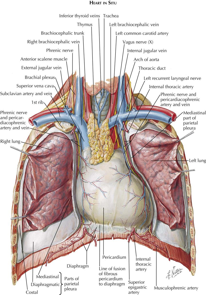
Organs & structures of the chest heart.
The chest is made up primarily of two muscles: Computed tomography (ct) of the chest can detect pathology that may not show up on a conventional chest radiograph(1). Swensen fund for innovation in teaching. The chest wall is comprised of skin, fat, muscles, and the thoracic skeleton. The myotomes elongate and invade the mesoderm of the wall of the embryonic thoracic and abdominal cavities. Anatomically, the heart is located in the anterior thoracic cavity; Related posts of anatomy of the chest and stomach anatomy of human body organs. A heart attack results from blocked blood flow, often from a blood clot, to your heart muscle. About the 6th week, the somites differentiate into the sclerotomes and the dermatomyotomes. The circulatory system does most of its work. It is important to remember the position and orientation of the heart when placing a stethoscope on the chest of a patient and listening for heart sounds, and also when looking at images taken from a midsagittal perspective. The right side of the heart is deflected anteriorly, and the left side is deflected posteriorly. In insects, crustaceans, and the extinct trilobites, the thorax is one of the three main divisions of the creature's body, each of which is in turn composed of multiple segments.
Chest pain has many possible causes, all of which need medical attention. These myotomes divide into the epimere and the hypomere. First i'll do an intro to the different organs and structures in the chest, and then i'll go over some images showing their locations. Related posts of anatomy of the chest area abdominal anatomy organs in quadrants. The chest is made up primarily of two muscles:
1 Anatomy Thoracic Key from thoracickey.com Chest a man's chest — like the rest of his body — is covered with skin that has two layers. The chest is the area of origin for many of the body's systems as it houses organs such as the heart, esophagus, trachea, lungs, and thoracic diaphragm. Download my two educational text books for free using this link: Swensen fund for innovation in teaching. The epidermis is the outermost layer that provides a protective, waterproof seal over the body. A heart attack results from blocked blood flow, often from a blood clot, to your heart muscle. The circulatory system does most of its work. Browse 2,549 female chest anatomy stock photos and images available, or start a new search to explore more stock photos and images.
A line is drawn from anterior surface of the body of 6th thoracic vertebrae passing through the apex of the heart up to anterior lower most part of diaphragm.
Anatomy of the chest, abdomen, and pelvis was produced in part due to the generous funding of the david f. Swensen fund for innovation in teaching. The dominant muscle in the upper chest is the pectoralis major. This tutorial is designed to help you understand the normal anatomy of the chest as seen on ct images in three planes: Angina is the term for chest pain caused by poor blood flow to the heart. The chest is the area of origin for many of the body's systems as it houses organs such as the heart, esophagus, trachea, lungs, and thoracic diaphragm. Learn about each of these muscles, their locations, functional anatomy and exercises for them. Plus, how to target each to make them bigger and stronger. The chest is made up primarily of two muscles: Diseases of the chest and chest abnormalities make up a significant portion of a physician's daily practice. Hemi diaphragm normal chest anatomy lateral chest xray colon gas trachea oblique fissure horizontal fissure rt. Learn vocabulary, terms, and more with flashcards, games, and other study tools. An overview of the anatomy visible in a transverse computed axial tomographical image of the thorax (and part of the abdomen) performed with intravenous cont.
Anatomy of human body organs 12 photos of the anatomy of human body organs anatomy human body organ systems foldable, anatomy of human body and its functions, anatomy of human body drawing pdf, anatomy of human body parts, anatomy of human body quiz, human anatomy, anatomy human body organ systems foldable. Related posts of anatomy of the chest and stomach anatomy of human body organs. Of the two chest muscles, the pectoralis major (a.k.a. This page provides an overview of the chest muscle group. It is important to remember the position and orientation of the heart when placing a stethoscope on the chest of a patient and listening for heart sounds, and also when looking at images taken from a midsagittal perspective.
Lateral Anatomy Of The Chest Abdomen And Bones Medical Art Works from cdn.shopify.com Saturday, june 5 from 4pm to 5pm pdt Anatomy of human body organs 12 photos of the anatomy of human body organs anatomy human body organ systems foldable, anatomy of human body and its functions, anatomy of human body drawing pdf, anatomy of human body parts, anatomy of human body quiz, human anatomy, anatomy human body organ systems foldable. The chest is the area of origin for many of the body's systems as it houses organs such as the heart, esophagus, trachea, lungs, and thoracic diaphragm. Organs & structures of the chest heart. These myotomes divide into the epimere and the hypomere. This page provides an overview of the chest muscle group. It provides protection to vital organs (eg, heart and major vessels, lungs, liver) and provides stability for movement. In insects, crustaceans, and the extinct trilobites, the thorax is one of the three main divisions of the creature's body, each of which is in turn composed of multiple segments.
Swensen fund for innovation in teaching.
Related posts of anatomy of the chest and stomach anatomy of human body organs. (1) the pectoralis major, and (2) the pectoralis minor. The muscles of the chest develop from the somites found in the mesoderm. About the 6th week, the somites differentiate into the sclerotomes and the dermatomyotomes. Pacemaker diagram cross section of a human heart with pacemaker fitted, showing the major arteries and veins. Of the two chest muscles, the pectoralis major (a.k.a. The chest is the area of origin for many of the body's systems as it houses organs such as the heart, esophagus, trachea, lungs, and thoracic diaphragm. Related posts of anatomy of the chest area abdominal anatomy organs in quadrants. Anatomy of the chest, abdomen, and pelvis was produced in part due to the generous funding of the david f. The chest wall is comprised of skin, fat, muscles, and the thoracic skeleton. Chest pain has many possible causes, all of which need medical attention. Plus, how to target each to make them bigger and stronger. It provides protection to vital organs (eg, heart and major vessels, lungs, liver) and provides stability for movement.


0 Comments:
Posting Komentar